
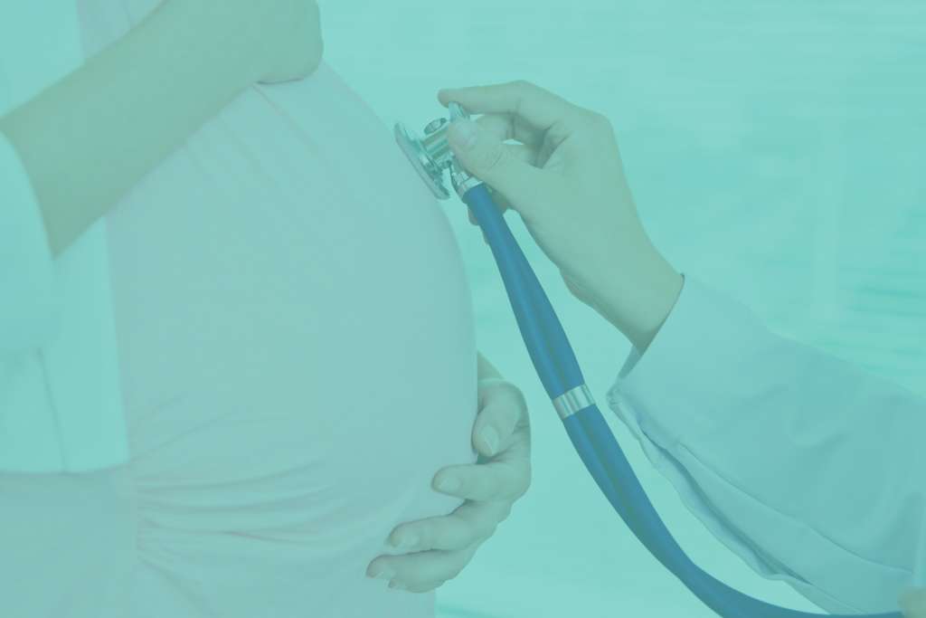
3D and 4D ultrasonography is a prenatal examination available in our gynaecological office, which allows to visualize in a spatial way, in real time, the image of the fetus. An advanced ultrasound device gives the possibility to see the fetus’ movements during the examination, and the image generated during the examination is three-dimensional. Currently, thanks to the 3D and 4D ultrasound examination, we have gained the possibility to perform a very reliable and extremely accurate diagnosis, but we also get the first, three-dimensional picture and video documenting the movements of our child. All this in digital format.
3D and 4D ultrasound is performed using the high-end and generation GE VolusonE8 Expert BT 2016 (the latest model available on the market):
- 3D/4D vaginal probe
- 3D/4D abdominal probe
Prenatal examination of the first trimester
This test is recommended by the Polish Gynaecological Society, so every pregnant woman should do it.
The optimal time to perform an ultrasound is 12 weeks of pregnancy (between 11 and 14 weeks). This is when the fetus is most optimally placed to assess nasal bone (NB) and neck translucency (NT) – and these are the most important genetic markers during this period of pregnancy. Apart from the markers mentioned above, we also assess the blood flow in the venous tract (DV) and through the tricuspid valve (TR), fetal heart function (FHR), parietal-socket length (CRL) and the fetus’ structure (skull, abdominal wall, stomach, bladder, spine, hands and feet).
The ultrasound in the first trimester of pregnancy can be extended by the so-called PAPP-A test, or biochemical test. It helps to increase diagnostic confidence in the fetus’ health.
Prenatal test of the second trimester
So-called half ultrasound (performed between 18 and 20 weeks of pregnancy).
Thanks to it, the doctor excludes genetic defects and assesses the correctness of the fetal anatomy. Its location, location and maturity of the placenta and the amount of fetal water are taken into account.
Examination of fetal anatomy includes the assessment of the shape and dimensions of the skull, brain, face, chest, heart, diaphragm, stomach, liver, kidneys, bladder of the umbilical cord trailer, and the continuity of the abdominal wall. Additionally, the spine, long bones and the structure of the fetus’ feet and hands are assessed.
Prenatal tests of the 2nd trimester are screening tests. They do not give 100% certainty of giving birth to a healthy child, but they allow to diagnose or exclude numerous birth defects.
NOTE! The quality of the fetal ultrasound image in 2D and 3D/4D technique and the time of the examination depends on many factors, among others:
- fetal arrangements
- gestational age
- the quantity of foetal water
- the amount of adipose tissue,
- application of abdominal skin creams before the test
Third trimester prenatal examination
Fetal ultrasound performed between 28th and 32nd week of pregnancy. It is helpful to assess the maturity of the fetus as well as detect any abnormalities that may have occurred after the second trimester ultrasound.
Ultrasound, which we perform in the last trimester of pregnancy, is important not only for the assessment of peripheral circulation condition or fetal development. It is also necessary to exclude that there are additional abnormalities that were not yet visible in previous studies.


