Obstetrics

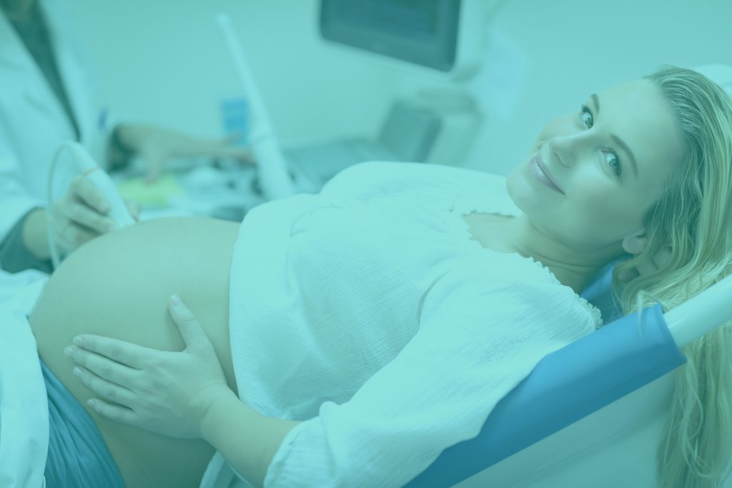
Ultrasound examination during pregnancy up to 10 weeks.

Ultrasound examination during pregnancy up to 10 weeks with a high resolution 3D/4D camera with description and exact calculation of the date of delivery.

The first visit to a pregnant gynaecologist should take place between 6 and 8 weeks. The doctor carries out a detailed interview and basic gynaecological and obstetric examination. During this visit the actual size of the pregnancy is determined and the date of birth from the last period is calculated and compared with the length of the embryo (USG). The fetus’ heartbeat, the so-called FHR, is also assessed. The doctor will also discuss the question of the required vitamin supplementation and the scope and forms of permitted physical activity. The gynaecologist then directs the patient to the midwife in order to establish the pregnancy card and determine what tests she should perform. The tests are ordered in accordance with the currently binding Standard of Peripheral Care.

Maternity visit during pregnancy

A normal, uncomplicated pregnancy requires about 10-12 visits every 4 weeks in early pregnancy, and every 3 weeks in the third trimester, in the last month even weekly, combined with a cardiotocographic record (fetal heart rate and uterine systolic function examination or flow Doppler ultrasound). More or less this is it: 5-6 weeks of pregnancy, 9 weeks, 12 weeks, 16 weeks, 20-22 weeks, 25-26 weeks, 30 weeks, 33 weeks, 36 weeks, 38 weeks, 39 weeks, 40 weeks

Before the visit, the patient goes to the midwife’s office, where she is weighed and her blood pressure is measured. The measurements together with the results of laboratory tests brought by the patient are recorded in the pregnancy card.

Then the gynaecologist-obstetrician is consulted. Each time the quantity and quality of vaginal discharge, its pH and the condition of the cervix are assessed. Depending on the situation, additional diagnostics is implemented – cytological smear, cervical canal culture.

During a follow-up obstetric visit, the presence of a normal fetal heart rate is always confirmed – usually by means of an ultrasound or a special fetal heart rate monitor.

During each maternity visit, a partner can accompany the future mother.

All kinds of certificates (e.g. for an employer with information about pregnancy) are issued at the patient’s request. If the patient’s state of health prevents her from doing her job, a L4 medical certificate is issued.

Test PAPP-A

The ultrasound and trimester can be extended to include the PAPP-A test, a non-invasive prenatal test. It consists in biochemical analysis of patient’s blood for the level of PAPP-A and free beta hCG.

The combination of biochemistry (PAPP-A test) and prenatal ultrasound allows for greater detection of chromosome aberrations, including Down’s syndrome, in the fetus.

In the case of the PAPP-A test, it is very important to perform it on specialist equipment compatible with the Fetal Medicine Foundation software, thanks to which the level of reliability of results is sufficiently high. In our office we work in the above mentioned program.

SANCO prenatal genetic test

The SANCO test is the only non-invasive, genetic prenatal test performed entirely in Poland. It determines the risk of chromosome trisomy of 21, 18 and 13 fetuses (Down’s, Edwards’ and Patau’s syndromes), as well as several other congenital defect syndromes.

The indication to perform the test is any suspicion of fetal genetic abnormalities resulting from, among others, pregnancy over 35 years of age, abnormal ultrasound results or in vitro fertilization.

The test requires only a small sample of future mother’s blood, whose plasma contains the child’s genetic material (so-called extracellular, fetal DNA, cffDNA). The detection rate of the test exceeds 99%.

Thanks to the high sensitivity and accuracy of the test, it allows to reduce the number of women for whom invasive testing is necessary six times.

DLACZEGO SANCO?

There are currently many possibilities for prenatal tests. However, compared to non-invasive prenatal genetic testing such as SANCO, traditional screening methods have a low sensitivity and a high percentage of false positives. Invasive methods such as amniocentesis or trophoblast biopsy (CVS) are reliable but carry a 0.5-2% risk of miscarriage.

Sanco Test RHD

It is made from the mother’s blood to assess the fetus’ RhD factor during pregnancy. This test is useful when qualifying a pregnant woman for intrapregnancy serological conflict prevention:

  • in case of complications
  • treatments
  • pregnant

In order to perform the test, a small amount of future mother’s blood is needed, whose plasma contains the child’s genetic material. Based on DNA analysis, the fetus is checked for RhD. The detection rate is over 99.5%. The test can be carried out between 12 and 24 weeks of pregnancy.

KTG – fetal heart record

The KTG examination, i.e. cardiotocography, consists in monitoring the fetus’ heart function and uterine muscle contraction. This examination allows us to detect possible abnormalities in the fetus’s development related to heartbeat disorders. KTG is performed in advanced pregnancy and during labour.

During the examination, two sensor belts are placed on the woman’s abdomen, which are connected to the monitor. The first belt measures the fetus’ heartbeat and the second one records the uterine contractions. The examination usually takes about 30 minutes, but in some cases it can take up to an hour.

NACE – Non-invasive prenatal test

NACE is a modern, non-invasive prenatal test. Using it we are able to diagnose most of the chromosomal abnormalities most commonly found in children (e.g. Down’s, Edwards’ and Patau’s syndrome, abnormal amount of sex chromosomes). It consists of taking a small amount of blood from the mother, from which free fetal DNA (so-called cffDNA) is isolated.

This test is completely safe for the mother and foetus. The test was developed by the American company Illumina, Inc. It is not only safe, but also extremely effective in diagnosing abnormalities resulting from excess or shortage of chromosomes.

The NACE test is available in 2 versions:

  • NACE Standard (Down, Edwards and Patau syndrome, fetal sex and sex chromosome changes – Turner and Klinefelter syndrome)
  • NACE Extended 24 (NACE Standard + additional 6 microdelegations: 22q11.2 (DiGeorge syndrome), 1p36, 15q11.2 (Angelman syndrome), 15q11.2 (Prader-Willi syndrome), 5p- (Cri du chat syndrome), 4p- (Wolf-Hirschhorn syndrome) and all chromosomes analysis

USG

Breast ultrasound

It facilitates the detection of pathological changes in the breasts – both benign and malignant. It is safe and can be repeated at will, unlike mammography.

It is recommended that breast ultrasound is performed prophylactically once a year in women under 40 years of age. In turn, in the case of older people, it can be a supplement to the mammogram.

Breast ultrasound is also performed when the doctor or the patient herself has noticed any nodules or other thickening, changes in the nipples or leaks in the nipples. The examination is performed with a high resolution camera for 3D/4D tests

Transvaginal ultrasound

This is a basic diagnostic method in modern gynaecology. It allows to assess the structures of both ovaries, mucous membrane (endometrium) and cervix.

It is much more accurate than an examination through the abdominal wall and can detect tumors, myomas, cysts or other small pelvic lesions. It is also the basic method of detecting polycystic ovarian syndrome.

It is made with a high-resolution GE Voluson E8 Expert camera with 3D/4D and Doppler imaging capabilities (vascular flow examination).

Gynecological ultrasound can be performed at any time in the cycle. It usually lasts several minutes and is not painful, but in some women it causes some discomfort.

Transvaginal ultrasound is performed as a basic element of gynaecological examination and is also an ultrasound examination in the first trimester of pregnancy.

.

ginekolog we wrocławiu , położnik , diagnostyka prenatalna wrocław badania prenatalne

Prenatal tests

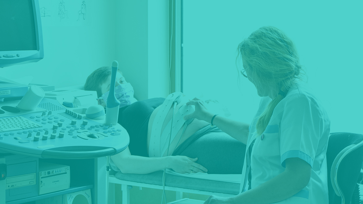
3D and 4D ultrasonography is a prenatal examination available in our gynaecological office, which allows to visualize in a spatial way, in real time, the image of the fetus. An advanced ultrasound device gives the possibility to see the fetus’ movements during the examination, and the image generated during the examination is three-dimensional. Currently, thanks to the 3D and 4D ultrasound examination, we have gained the possibility to perform a very reliable and extremely accurate diagnosis, but we also get the first, three-dimensional picture and video documenting the movements of our child. All this in digital format.

3D and 4D ultrasound is performed using the high-end and generation GE VolusonE8 Expert BT 2016 (the latest model available on the market):

  • 3D/4D vaginal probe
  • 3D/4D abdominal probe
Prenatal examination of the first trimester

This test is recommended by the Polish Gynaecological Society, so every pregnant woman should do it.

The optimal time to perform an ultrasound is 12 weeks of pregnancy (between 11 and 14 weeks). This is when the fetus is most optimally placed to assess nasal bone (NB) and neck translucency (NT) – and these are the most important genetic markers during this period of pregnancy. Apart from the markers mentioned above, we also assess the blood flow in the venous tract (DV) and through the tricuspid valve (TR), fetal heart function (FHR), parietal-socket length (CRL) and the fetus’ structure (skull, abdominal wall, stomach, bladder, spine, hands and feet).

The ultrasound in the first trimester of pregnancy can be extended by the so-called PAPP-A test, or biochemical test. It helps to increase diagnostic confidence in the fetus’ health.

Prenatal test of the second trimester

So-called half ultrasound (performed between 18 and 20 weeks of pregnancy).

Thanks to it, the doctor excludes genetic defects and assesses the correctness of the fetal anatomy. Its location, location and maturity of the placenta and the amount of fetal water are taken into account.

Examination of fetal anatomy includes the assessment of the shape and dimensions of the skull, brain, face, chest, heart, diaphragm, stomach, liver, kidneys, bladder of the umbilical cord trailer, and the continuity of the abdominal wall. Additionally, the spine, long bones and the structure of the fetus’ feet and hands are assessed.

Prenatal tests of the 2nd trimester are screening tests. They do not give 100% certainty of giving birth to a healthy child, but they allow to diagnose or exclude numerous birth defects.

NOTE! The quality of the fetal ultrasound image in 2D and 3D/4D technique and the time of the examination depends on many factors, among others:

  • fetal arrangements
  • gestational age
  • the quantity of foetal water
  • the amount of adipose tissue,
  • application of abdominal skin creams before the test
Third trimester prenatal examination

Fetal ultrasound performed between 28th and 32nd week of pregnancy. It is helpful to assess the maturity of the fetus as well as detect any abnormalities that may have occurred after the second trimester ultrasound.

Ultrasound, which we perform in the last trimester of pregnancy, is important not only for the assessment of peripheral circulation condition or fetal development. It is also necessary to exclude that there are additional abnormalities that were not yet visible in previous studies.

ginekolog we wrocławiu , położnik , diagnostyka prenatalna wrocław ginekologia estetyczna

Aesthetic Gynaecology

Vaginal and vulva laser revitalization – Aphrodite laser

The births and natural aging processes cause changes in the female body. These changes affect both our physical and mental health, which affects self-esteem and satisfaction with life. Aesthetic gynaecology comes in handy, which uses minimally invasive procedures to treat illnesses and solve problems that have so far required surgical treatment.

Aphrodite is an innovative endovaginal (vaginal) laser used to reduce the symptoms of the following conditions:

  • Vaginal atrophy

A common condition caused by hormonal changes caused by estrogen deficiency. Symptoms: vaginal dryness, inflammation, pain during and bleeding after intercourse, burning, itching, decreased vaginal elasticity.

  • Vaginal loosening syndrome (VRS) and the associated reduced sexual experience

VRS occurs most often after birth and during the menopause. It consists in the loss of optimal vaginal structure and tension and is a serious problem in intimate contacts.

  • Labioplasty of vulva lips

Asymmetry or too big a size of vulva lips can be not only an aesthetic but also functional problem for many women. They can have an impact on lowering self-acceptance and, consequently, on problems with coexistence.

  • Effortless urinary incontinence (WNM)

This problem affects a significant proportion of the female population, usually related to childbirth or to the passing of time. The vaginal walls are lowered, which reduces the pressure of the vagina on the urethra and as a result of, for example, physical effort or coughing, more or less urine is passed.

Thanks to the use of Aphrodite laser it is possible to thoroughly revitalize the mucous membrane and thus restore the natural vaginal pH. The laser acting thermally on the vaginal mucous membrane makes it tighter and better hydrated, which directly increases the sexual satisfaction.

The Aphrodite laser also enables the reduction of external intimate organs, providing a shorter healing time compared to classical surgical methods and reducing the discomfort associated with the procedure. In case of stress urinary incontinence, the laser can also be helpful. Its action affects the contraction of vaginal walls, it also increases its tension and flexibility, which increases its pressure towards the urethra and bladder.

Procedure and effects

The treatment is painless and takes about 15 minutes. During the procedure, the doctor uses a laser beam, which he introduces into the vagina causing shrinkage and stimulation of collagen fibers, so that the vaginal wall is strengthened and stiffened. Also the position of the urethra is corrected, restoring normal functioning of the urinary system. By shrinking the uterine bottom muscles, normal functioning of the urinary system is restored and the woman regains self-confidence and comfort of living. Immediately after the procedure the patient can return to her daily activities.

Revitalization of the scars after Caesarean section / incision, perineal rupture with Aphrodite laser

After childbirth, there is often an abnormal healing process due to inaccurate stitching of the wound, or abnormal healing due to secondary infection or suture propagation.

Laser correction of the scar after perineal incision/fracture or Caesarean section is a minimally invasive procedure involving controlled micro-damage of scar tissue by means of laser rays, which initiates the process of collagen fiber production and its reconstruction. Collagen fibres become shorter and thicker, which makes the scar smooth, shallow and additionally brighten up.

The procedure lasts about 15, minutes, is performed under local anesthesia, the patient can go home immediately after the procedure.

Consultation of the midwife

Keeping a midwife pregnant at the NFZ

Any woman whose pregnancy is physiological has the right to be guided by a midwife at the NFZ. The midwife, in accordance with the regulations of the Minister of Health, has the competence and knowledge to lead the pregnancy on her own.

Why choose a midwife?

  • A midwife’s physiological pregnancy gives the woman the certainty of professional and friendly care.
  • The midwife will also ensure continuity of care as she accompanies her mother both during the pregnancy and takes care of her and the baby during the first weeks after the birth.

In our facility, the midwife leading the pregnancy is Jolanta Petersen, MA.

Standard midwife’s visit

  • During the first meeting: putting on a pregnancy card, recognizing the needs of the pregnant woman, ordering laboratory tests according to the Standard of Perinatal Care.
  • Subsequent meetings: supplementation and analysis of laboratory tests, control of pressure, weight, vaginal pH, fetal movements, auscultation of the fetus’s heart rate, external examination, discussion of emerging ailments and natural ways of alleviating them, discussion of the correct diet, supplementation and physical activity, advice on alleviating anxiety and anxiety related to pregnancy/childbirth.

With the midwife at the end of the pregnancy, the birth plan is completed together and KTG tests are performed.

The midwife is obliged to refer a woman to a gynaecologist three times during pregnancy for ultrasound prenatal tests and always in case of abnormalities.

In our clinic, ultrasound examinations are performed by Dr Tomasz Michalik – an outstanding specialist in carrying out ultrasounds of the first, second and third trimester as well as high risk pregnancies.

Visits to the midwife usually take place once a month, at the end of pregnancy more often every 2 weeks.

What else falls within the competence of a midwife?

PREVENTIONAL CARE – the midwife can provide information on how to prepare the pregnancy properly, what tests to perform, what to look out for in terms of diet, supplementation and physical activity.

Pregnancy care – the midwife conducts, among others, free educational meetings for pregnant women.

CONSUMPTION OF AIR CONDITION – the midwife can carry out and accept a physiological birth on her own, both in hospital and at home.

PATHERSONAL VISITS – a postpartum midwife can take professional care of a woman in childbirth and her newborn baby. The midwife should visit the mother and child in their home at least 4 times. You can count on the midwife’s support until the baby is two months old.

SUPPORT FOR FIRST-feeding – the midwife also has the knowledge and competence to support the woman in breastfeeding, help solve problems, give lactation advice and promote natural feeding.

And also:

  • Post-gynaecological or oncological-gynaecological surgery care
  • SUPPORT during the menopause
  • EDUCATION at every stage of life in the field of pro-healthy lifestyle, prevention of HIV infection and sexually transmitted diseases, cancer, obstetric pathologies.

Pre-natal education programme
led by midwife Jolante Petersen, MA.

  • The course of pregnancy and development of the child from the beginning of pregnancy to delivery, lifestyle during pregnancy – diet, professional activity, physical activity, ailments of the pregnancy period and ways of coping with them, prevention of infectious diseases in the perinatal period, psychological and emotional problems of a woman and her family during pregnancy, delivery and postpartum.
  • Factors that herald childbirth, or how to choose the right moment to come to the hospital? How to pack a bag for delivery? Where to give birth? – discussion of hospitals in Wrocław and its surroundings.
  • Physiology of childbirth, active childbirth, family childbirth; positions used in the first and second period of childbirth (vertical positions) and equipment helpful during childbirth; relieving labour pain (non-pharmacological and pharmacological); Is breathing important in childbirth? How to calm down during childbirth and focus on working with the body? What is a delivery cocktail and intervention cascade?
  • Individual preparation of the Birth Plan, discussion of preferences and expectations of the parturient.
  • Caesarean section – benefits and risks for mother and child, how to ensure quick contact with the baby after surgery, what is “baby seeding”?
  • Midwifery – the course, symptoms, return of fertility after birth, psychological aspects of the postpartum; Why are the pelvic floor muscles so important and why do we have to work with them? Who is a urogynological physiotherapist and why is it worth visiting him already 3-4 weeks after the birth?
  • Care of the newborn baby and then of the infant – how to take care of the umbilical cord stump, what to do with the abscessing eye? Baby care: instruction in proper wearing, changing, dressing, massage and first aid show. How to properly complete a layette for your baby?
  • Breastfeeding and lactation support, including troubleshooting; lactation hemorrhage, warts, food stasis, and breast inflammation. When is it worth contacting a lactation counsellor? Translated with www.DeepL.com/Translator (free version)

ginekolog we wrocławiu , położnik , diagnostyka prenatalna wrocław

Gynaecology

Gynaecological visit

A gynaecological visit starts with an interview with a doctor who asks about past diseases, pregnancies and other information he needs. It also includes a palpable breast examination. The gynaecological examination takes place on a gynaecological chair and consists in the evaluation of vulva, vaginal mucosa and cervix. Vaginal discharge is collected for microscopic evaluation. – The so-called direct smear (it can detect a fungal or bacterial infection of the vagina). Then, the transvaginal palpation test, the so called combined, is evaluated the uterus and appendages (ovary and fallopian tube).

Then the couch is used for transvaginal ultrasound with a high resolution camera, also used for 3d/4D tests (GE Voluson E8 Expert).Then the couch is used for transvaginal ultrasound with a high resolution camera, also used for 3d/4D tests (GE Voluson E8 Expert).

Comprehensive study

On one visit, I carry out comprehensive prophylactic tests, which include:

  • Gynaecological examination
  • Transvaginal gynaecological ultrasound
  • Palpation and breast ultrasound
  • Cytology

After the examination, its results are discussed and prescriptions are written. During the examination, it is also possible to collect cytological examination material.

Cytology

It is used for the prevention and diagnosis of pre-cancerous conditions as well as cervical cancer, which is one of the most common cancers in Poland. During the examination, the doctor or certified nurse takes a sample of exfoliated epithelium from the patient’s cervix with the help of a sight glass.

Cytological examination is not painful. It is recommended that the first time they are performed between twenty-five years of age or soon after the start of intercourse. This is a routine examination, which is usually performed once a year, during the gynaecological visit.

In our office, cytology can also be performed at the National Health Fund as part of the Cervical Cancer Prevention Programme. The programme is intended for women aged 25 – 59 (according to the yearbook) who have not performed a cytological examination within the last 3 years. The cytological material is evaluated in two centres at the DCO (Lower Silesian Oncology Centre) located at Hirszfeld Sq. or at the Histmed histopathological laboratory – Dr Kosiński. Each examination is verified by a histopathologist. If any disturbing changes are detected, the next examination which is recommended is a colposcopy.

Recommendations

  • The optimal period for the collection of cytological material is between 10 and 20 days of the cycle
  • Sexual intercourse must be refrained from at least 24 hours before the examination
  • For a few days prior to the examination, vaginal medications or treatments on the vagina and cervix should not be used.
  • Cytology may be performed at least one day after gynaecological or vaginal ultrasound examination
  • Smears for cytological examination should be taken at least 2 – 3 days after the end of menstrual bleeding
  • The examination in the sight glass should precede the examination through the vagina no – smears should be taken immediately after the gynaecological examination
  • No cytological examination shall be performed during active infection of the genital tract with associated discharge.
Liquid cytology

Also called thin layer, it is a modern technique of preparing cytological preparations. It allows for a more accurate diagnosis of HPV virus and cervical cancer. Preparation and storage of the taken sample differs from traditional cytology. In the LBC the brush with the material is immediately immersed in a special liquid. In it the swab is delaminated so that cells (e.g. bacteria) that may obscure the image of the sample being evaluated can be removed. This shows a much higher accuracy compared to traditional cytology when detecting both precancerous conditions and cancer cells. An important advantage is also the possibility to store cellular material (which is embedded in a liquid medium) and perform additional tests on the same sample (e.g. HPV).

Microbiological smear

The purpose of this examination is to detect and identify pathogens present in samples of biological material taken from the vagina or cervical canal. The result of the test indicates the existence or absence of vaginal inflammation and its possible type.

Test for HPV

The test allows to detect the DNA of 12 high-oncogenous genotypes of Human Papilloma Virus (HPV): 16, 18, 31, 33, 35, 39, 45, 51, 52, 56, 58, 59. These genotypes constitute almost all detected types of the virus which are responsible for cervical cancer. The Real Time – PCR method used in the study allows the detection of the virus genetic material with extremely high sensitivity and specificity. Detection of high-oncogenous HPV types in the patient gives the possibility to monitor possible changes and implement effective treatment.

Test for P16 protein

Intended for patients in whom the cytological examination resulted in ASCUS, LSIL or in other cases difficult to interpret (no unambiguous diagnosis can be made).

It is a complementary diagnosis of cervical cancer. It detects cancer at an early stage of its development. It identifies cells with a disturbed cell cycle, which may be caused by injection of highly ocogenous HPV. This test is an immunological test, which is based on monoclonal antibodies directed in cytological preparations against p16 protein. The p16 protein plays an essential role in controlling the normal cell cycle. In mature cells their level is significantly lowered (they are barely detectable), while in cervical cells, where there are already unfavorable changes, p16 levels are recorded as significantly elevated.

An additional advantage of the currently available latest version of the test is the presence of the second Ki-67 biomarker, i.e. cell proliferation protein (cell proliferation). Its application allows to obtain maximum sensitivity of the test. The test material is a smear from the cervical canal.

Colposcopy

An examination during which the doctor, using a specialist optical device, obtains a spatial image of the cervix, vulva and vagina. This allows the doctor to effectively assess the colour or structure of the epithelium for the diagnosis of diseases, in particular cervical cancer. Colposcopy is usually performed as a supplement to the cytological examination, in case of abnormal results.

During the examination, the cervix is washed with a special liquid to facilitate the observation of cell changes. If the image is abnormal, the doctor may also take a tissue sample for examination, which is then subjected to a histopathological examination. This examination allows you to make a diagnosis and determine the next steps of treatment.

It is recommended that a few days before the examination, vaginal irrigation and gynaecological examinations should be abandoned, as this may adversely affect the assessment of colposcopic image. Colposcopia is best reported after menstruation.

It is also advisable for the examination to be performed by a woman with the result of the last cytology, which was the indication for colposcopy.

Ovulation diagnostics

It consists in monitoring ovulation by means of ultrasound, e.g. to diagnose the causes of infertility.

During one cycle three ultrasounds are performed to determine the size of the follicles in the ovaries and to try to determine the moment of ovulation itself.

Venereal Package

The most common diseases of the urogenital system are chlamydiosis, mycoplasmosis, and gonorrhea. Ureaplasma infection and syphilis are also relatively common. The most important risk factors for urogenital infections are:

  • large number and/or frequent changes of sexual partners
  • use of oral contraceptives
  • antibiotic therapy
  • certain diseases, such as diabetes, cancer
  • a condom reduces the risk of contracting sexually transmitted diseases but does not eliminate it completely!

most of the above mentioned diseases do not give any symptoms at first, but later on they are easily confused with other disease entities, e.g. with an allergy (this is the case with syphilis, which initially causes a papular rash). Untreated infections lead to serious complications such as pelviculitis, urethritis, premature birth, developmental defects in the unborn child (low birth weight, neurological disorders) and infertility.

Cervical cryotherapy

A method otherwise known as “freezing”. Used to remove benign lesions that are located within the cervix. It is effective and almost painless – it does not require an anaesthetic. Diseased cells (erosions) are destroyed by low temperature (-192 degrees Celsius). Freezing with liquid nitrogen does not damage and thus does not cause functional disorders of the cervix. However, it is not possible to take the material for histopathological examination. Requirements for the procedure: correct cytological examination result, colposcopic examination result, first phase of the cycle. The procedure takes about 20-30 minutes.

Endometrial aspiration biopsy

It’s a minimally invasive procedure that involves taking a piece of uterine mucosa. It involves inserting a thin, sterile tube into the uterine cavity (through the cervical canal) and collecting pieces of uterine mucosa by creating a vacuum. The whole procedure takes about a few dozen seconds. The collected material will then be examined under a microscope.

Indications for this test are intermenstrual and postmenopausal spotting and bleeding, hormonal fluctuations, problems with getting pregnant.

Aspiration biopsy of the endometrium is a very effective diagnostic tool, and due to its safety and ease of technical performance, it allows to pre-diagnose or exclude disturbing proliferative changes in the endometrium, including endometrial cancer. According to the studies carried out so far to assess the effectiveness of this method, this test is as effective as curettage of the uterine cavity.

Domestic insemination

It’s a minimally invasive procedure that involves taking a piece of uterine mucosa. It involves inserting a thin, sterile tube into the uterine cavity (through the cervical canal) and collecting pieces of uterine mucosa by creating a vacuum. The whole procedure takes about a few dozen seconds. The collected material will then be examined under a microscope.

It is a safe and painless procedure involving placing a sample of semen in the uterus, which was previously properly prepared in the laboratory.

The intrauterine insemination procedure can be used by couples who:

  • they don’t have a baby, despite correct test results and long efforts to get pregnant
  • suffer from idiopathic infertility
  • have at their disposal semen that is slightly abnormal (insufficiently mobile and not very numerous sperm);
  • they experience ejaculation disorders (patient) or are found to have endometriosis (1st and 2nd degree) or are found to have sperm antibodies (patient).

The insemination procedure can be repeated many times – to the end – but usually no more than six tests are performed in consecutive monthly cycles. Depending on the age of the patient, the quality of semen and the number of mature follicles after hormonal stimulation, the effectiveness of intrauterine insemination is between 10 and 25% per cycle.